Description
Clinical History
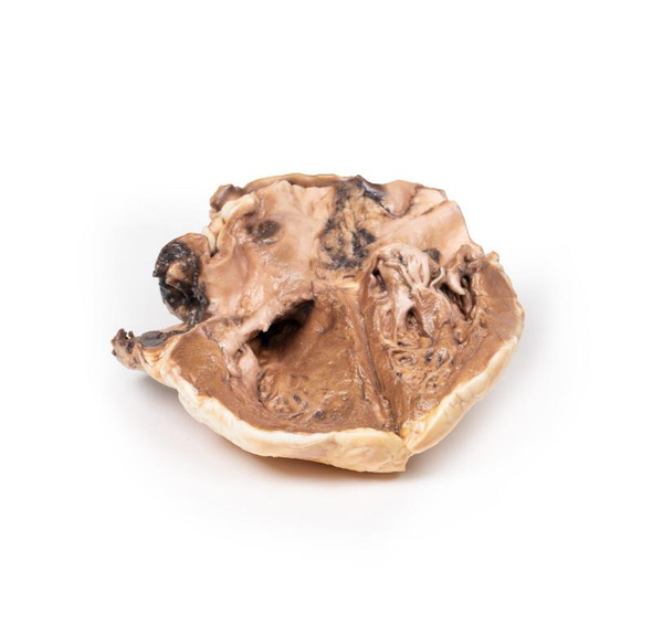
A 68-year-old female presented with recent onset of seizures and was diagnosed with epilepsy. Collateral history revealed a gradual change in the patient's personality. She subsequently died several months later from a myocardial infarction.
Pathology
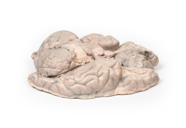
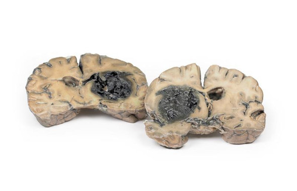
This brain specimen has been sliced horizontally. A well-circumscribed 6cm tumor is evident in between the two frontal lobes. The tumor is compressing the frontal lobes. It has a pinkish cut surface with some yellow areas indicating necrosis. It was attached to the dura mater anteriorly. This is an example of a meningioma.
Further Information
Meningiomas are often said to be the most common tumors of the central nervous system (CNS); however, in fact they arise in the meninges (dura, arachnoid and pia), which are strictly speaking not part of the CNS per se. They arise from arachnoid cells closely associated with the dura; hence, these tumors can be associated with the dura or dural folds (falx cerebri and tentorium cerebelli). Meningiomas are predominantly slow growing benign tumors. Symptoms are determined by the tumor location and the speed of growth. Symptoms include seizures, change of mental state, vision, hearing- or smell alterations, and symptoms of increased intracranial pressure. Meningiomas are frequently asymptomatic. Treatment includes observation, surgery or radiotherapy, depending on the clinical context and tumor morphology.
Meningiomas are rare in children with a median age of 65 years at diagnosis. There is a 3:2 female predominance. Exposure to ionizing radiation, including cranial radiotherapy, increases the risk of development meningiomas. The greatest genetic predisposition for development is seen in patients with neurofibromatosis type 2 (NF2). NF2 is an autosomal dominant disease caused by mutations in the NF2 gene on Chromosome 22 leading to multiple tumors associated with the nervous system.
Advantages
- Anatomically accurate and identical to real specimen
- No ethical issues - not real human body parts
- Reasonably priced
- Available within a short lead time
- Reproducible, several identical prints can be used as a classroom set
- Can be produced in different sizes to cater for the needs of the teacher
Human Cadavers
- Access to cadavers can be problematic. Many countries cannot access cadavers for cultural and religious reasons
- Cadavers cost a lot money
- High cost for establishing your own plastination suite
- Wet specimens cannot be used in uncertified labs
- Dissection of cadavers is a lot of staff time and that is a cost
- Storage of cadaver material needs special refrigeration etc. which has coast
- If you want another specimen you have to start all over again
Plastinates
- Costs
- Ethical issues
- Timeframe for plastination process
- Many countries do not allow their importation
- One of a kind
Superior 3D print results compared with conventional methods
- Vibrant color offering with 10 million colors
- UV-curable inkjet printing
- High quality 3D printing that can create products that are delicate, extremely precise and incredibly realistic
Clear Support Material
- To avoid breakage of fragile, thin, and delicate arteries, veins or vessels, a clear support material is printed on such spots. This makes the models robust and can be handled by students easily.


