Description
Clinical History
No clinical details are available for this specimen.
Pathology
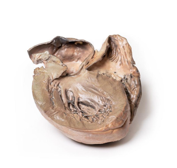
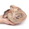
The heart displays both ventricles from the posterior aspect. There is a prominent saccular dilatation of the thoracic ascending aorta, which shows several atherosclerotic plaques and posteriorly is seen to be ruptured (identified by the dark staining). Both ventricles are hypertrophied. The coronary arteries together with the aortic and tricuspid valves are normal. This is an example of a ruptured aneurysm of the ascending aorta.
Further Information
The dilation of the ascending aorta is a common incidental finding on transthoracic echocardiography performed for unrelated indications.
The thoracic aorta is divided into 3 parts: ascending, arch and descending. The ascending aorta originates beyond the aortic valve and ends right before the innominate artery (brachiocephalic trunk). It is approximately 5 cm long and is composed of two distinct segments. The lower segment, known as the aortic root, encompasses the coronary sinuses and sinotubular junction (STJ). The upper segment, known as the tubular ascending aorta, begins at the STJ and extends to the aortic arch (innominate artery). More than 50% of thoracic aortic aneurysms are localized to the ascending aorta, which may affect either the aortic root or tubular aortic segment.
An aneurysm is defined as a localized dilation of the aorta that is more than 50% of predicted (ratio of observed to expected diameter = 1.5). Aneurysm should be distinguished from ectasia, which represents a diffuse dilation of the aorta less than 50% of normal aorta diameter. The incidence of ascending thoracic aortic aneurysms is estimated to be around 10 per 100,000 person-years[1].
Reference
1. Saliba et al. (2015). Int J Cardiol Heart Vasc. 6
Advantages
- Anatomically accurate and identical to real specimen
- No ethical issues - not real human body parts
- Reasonably priced
- Available within a short lead time
- Reproducible, several identical prints can be used as a classroom set
- Can be produced in different sizes to cater for the needs of the teacher
Human Cadavers
- Access to cadavers can be problematic. Many countries cannot access cadavers for cultural and religious reasons
- Cadavers cost a lot money
- High cost for establishing your own plastination suite
- Wet specimens cannot be used in uncertified labs
- Dissection of cadavers is a lot of staff time and that is a cost
- Storage of cadaver material needs special refrigeration etc. which has coast
- If you want another specimen you have to start all over again
Plastinates
- Costs
- Ethical issues
- Timeframe for plastination process
- Many countries do not allow their importation
- One of a kind
Superior 3D print results compared with conventional methods
- Vibrant color offering with 10 million colors
- UV-curable inkjet printing
- High quality 3D printing that can create products that are delicate, extremely precise and incredibly realistic
Clear Support Material
- To avoid breakage of fragile, thin, and delicate arteries, veins or vessels, a clear support material is printed on such spots. This makes the models robust and can be handled by students easily.


