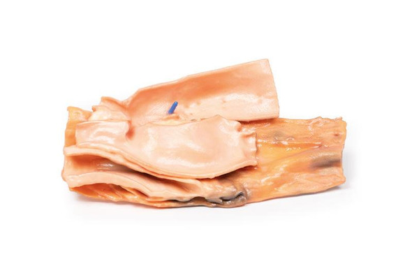
Description
Clinical History
This was a case of a 75-year-old female who presented with symptoms of recurrent disease and confirmed to have chemo-resistant multiple retroperitoneal lymph node metastases five years after the initial therapy for stage IIIc serous adenocarcinoma of the ovary. Positron emission tomography/computed tomography (PET/CT) revealed the involvement of para-aortic nodes and pelvic nodes. She died of liver complications before therapeutic options, such as radical lymphadenectomy, could be considered.
Pathology
The specimen consists of the abdominal aorta and common iliac arteries surrounded by large numbers of extremely enlarged iliac nodes para-aortic lymph nodes. Histopathological examination revealed metastatic high-grade adenocarcinoma in some of the resected lymph nodes.
Further Information
Occasionally, lymph node metastases represent the only component at the time of recurrence of ovarian cancer. In this case, the metastatic nodes predicted by PET/CT completely corresponded to the actual metastatic nodes. Ultrasound (US) could equally have confirmed the presence of such large lymph nodes. PET/CT or US often fails to identify microscopic disease in histopathologically-proven positive nodes. Therefore, it is difficult to reliably exclude lymph node metastases during surveillance following initial surgery for ovarian cancer.
In the context of a recurrent ovarian disease, systematic aortic and pelvic node dissection would be considered appropriate in younger women with no other evidence of metastatic disease. This is unlikely to be curative but may produce palliation with symptom control and allow for the trial of novel therapies if available.
More commonly, sampling of pelvic and aortic lymph nodes is part of the formal surgical staging of epithelial ovarian cancer at the time of initial surgical treatment. In addition, in women presenting initially with advanced stage ovarian cancer, systematic debulking of enlarged retroperitoneal lymph nodes will be considered if this leads to complete debulking of the tumor.
Advantages
- Anatomically accurate and identical to real specimen
- No ethical issues - not real human body parts
- Reasonably priced
- Available within a short lead time
- Reproducible, several identical prints can be used as a classroom set
- Can be produced in different sizes to cater for the needs of the teacher
Human Cadavers
- Access to cadavers can be problematic. Many countries cannot access cadavers for cultural and religious reasons
- Cadavers cost a lot money
- High cost for establishing your own plastination suite
- Wet specimens cannot be used in uncertified labs
- Dissection of cadavers is a lot of staff time and that is a cost
- Storage of cadaver material needs special refrigeration etc. which has coast
- If you want another specimen you have to start all over again
Plastinates
- Costs
- Ethical issues
- Timeframe for plastination process
- Many countries do not allow their importation
- One of a kind
Superior 3D print results compared with conventional methods
- Vibrant color offering with 10 million colors
- UV-curable inkjet printing
- High quality 3D printing that can create products that are delicate, extremely precise and incredibly realistic
Clear Support Material
- To avoid breakage of fragile, thin, and delicate arteries, veins or vessels, a clear support material is printed on such spots. This makes the models robust and can be handled by students easily.


