Description
Clinical History
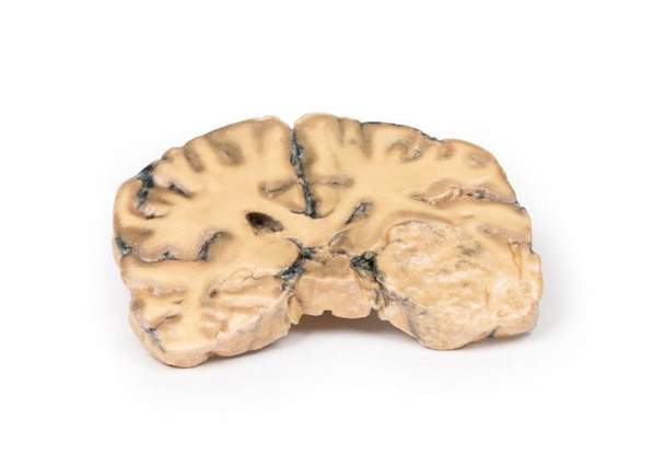
A 73-year-old female was admitted with new left-sided hemiplegia. On further questioning she revealed a 3-month history of headaches, nausea and deteriorating balance. CT brain revealed an inoperable brain tumor. She died 1 week after being admitted.
Pathology
This brain specimen is a coronal section. In the right temporal lobe, a poorly demarcated tumor is present. There is enlargement of the hemispheres and flattening of the gyral pattern. From the posterior aspect of the specimen subfalcine herniation is appreciated and the tumor appear less well differentiated with hemorrhagic and necrotic foci. Histology of this tumor showed an astrocytoma, Grade III/IV.
In subfalcine (or cingulate) herniation, the most common type of brain herniation, the innermost part of the frontal lobe is pushed under part of the falx cerebri, between the two hemispheres of the brain.
Further Information
Gliomas are the second most common cancer of the central nervous system after meningiomas. The term glioma refers to tumors that are histologically similar to normal glial cells i.e. astrocytes, oligodendrocytes and ependymal cells. They arise from a progenitor cell that differentiates down one of the cell lines. Astrocytomas develop from the astrocyte lineage of glial cells. Tumors are staged according to histological differentiation and range from diffuse astrocytoma (Grade II/IV) to anaplastic astrocytoma (Grade III/IV) to glioblastoma (Grade IV). Histological features include the prominent eosinophilic cytoplasm in some astrocytic tumor cells (gemistocytes) as well as a fibrillary background.
Astrocytomas occur most commonly between the fourth and sixth decades of life. Tumors usually occur in the cerebral hemispheres but may also occur in the cerebellum, brainstem or spinal cord. They most commonly present with seizures, headaches, nausea and focal neurological deficits depending on area involved. Without treatment Grade III median survival is 18 months. Treatment includes surgical resection, radiotherapy, chemotherapy or a combination thereof, depending on the clinical context.
Advantages
- Anatomically accurate and identical to real specimen
- No ethical issues - not real human body parts
- Reasonably priced
- Available within a short lead time
- Reproducible, several identical prints can be used as a classroom set
- Can be produced in different sizes to cater for the needs of the teacher
Human Cadavers
- Access to cadavers can be problematic. Many countries cannot access cadavers for cultural and religious reasons
- Cadavers cost a lot money
- High cost for establishing your own plastination suite
- Wet specimens cannot be used in uncertified labs
- Dissection of cadavers is a lot of staff time and that is a cost
- Storage of cadaver material needs special refrigeration etc. which has coast
- If you want another specimen you have to start all over again
Plastinates
- Costs
- Ethical issues
- Timeframe for plastination process
- Many countries do not allow their importation
- One of a kind
Superior 3D print results compared with conventional methods
- Vibrant color offering with 10 million colors
- UV-curable inkjet printing
- High quality 3D printing that can create products that are delicate, extremely precise and incredibly realistic
Clear Support Material
- To avoid breakage of fragile, thin, and delicate arteries, veins or vessels, a clear support material is printed on such spots. This makes the models robust and can be handled by students easily.


